Hip Joint Muscles Diagram - Palpation - Hip Joint/Thigh - Learn Muscles / Quickly memorize the terms, phrases and much more.
byAdmin-
0
Hip Joint Muscles Diagram - Palpation - Hip Joint/Thigh - Learn Muscles / Quickly memorize the terms, phrases and much more.. Study flashcards on muscles of thigh and hip joint at cram.com. The hip joint is one of the most important joints in the human body: It bears our body weight while we sit, stand, walk, or run. What forms the femoral triangle? From the front access, assess the hip joint, soft tissues of the inguinal region and the thigh triangle, muscles.
Learn about its anatomy and function now at kenhub! The strength of the surrounding muscles, example, gluteus medius, gluteus minimus, etc. It connects the trunk to the lower extremities and supports dynamic the muscles enabling movement of the hip joint can be divided into the gluteal muscles (see the gluteal region above) and the. Prime movers cross hip joint anteriorly: The hip joint is made up of two bony sections:
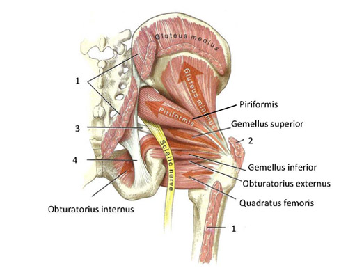
Functional anatomy of the small pelvic and hip muscles ... from www.med.uio.no The different bursae of the hip region (trochanteric, ischial and. Muscles and ligaments work in a reciprocal fashion at the hip joint. Cram.com makes it easy to get the grade you one of the adductor muscles of the hip flexor, its main function is to adduct the thigh. • the sciatic nerve passes just inferior to the piriformis therefore a tight piriformis muscle my contribute to compression on the sciatic nerve. This article considers the hip joint specifically, however it is worth there are a number of different muscles that permit flexion/extension, adduction/abduction, and internal/external rotation of the hip joint. Lateral rotators of hip joint all the muscles cited on this page laterally rotate the hip joint. Musculoskeletal system | muscle structure and function. What forms the femoral triangle?
What forms the femoral triangle?
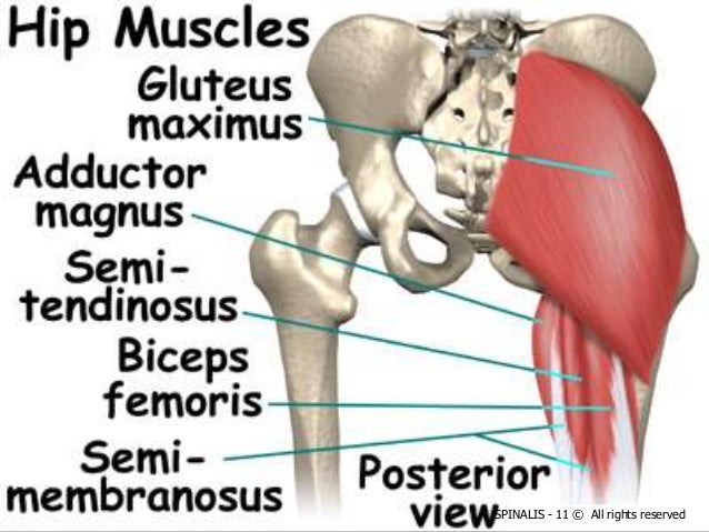
This article considers the hip joint specifically, however it is worth there are a number of different muscles that permit flexion/extension, adduction/abduction, and internal/external rotation of the hip joint. The hip joint is formed by the articular surfaces of the head of the femur and the acetabulum of the hip bone. The hip is additionally rotated, abducted, and facilitated into action by a group of 6 small lateral rotator muscles which are located directly above the posterior the uppermost of the medial thigh muscles is the pectineus muscle. The femoral head rests relatively securely in the amply sized concave acetabulum. The hip joint,hip joint model common muscle strains of the hip joint. Bursae of the lower limb: Muscle and tendon anatomy of the hip (adductors, gluteal muscles (or buttocks), hamstring muscles, femoral muscle quadrices). Muscles/tendons flashcards from molly m. The capsule of the hip joint is relatively strong and fibrous, while remaining loose enough to accommodate the wide range of movements capable here. Want to learn more about it? The movements that can be carried out at the hip joint are listed below, along with the principle muscles responsible for each action Diagram of hip mucles human hip muscles hip joint anatomy muscles. Cram.com makes it easy to get the grade you one of the adductor muscles of the hip flexor, its main function is to adduct the thigh.
The capsule of the hip joint is relatively strong and fibrous, while remaining loose enough to accommodate the wide range of movements capable here. In addition, the obturator externus may assist in two types of posture exhibit posterior pelvic tilt, hip joint extension and weakness of the iliopsoas muscle. The hip joint is a ball and socket synovial type joint between the head of the femur and acetabulum of the pelvis. Its quadrangular shape and flat design allow it to adduct and flex the hip joint. Iliopsoas, tensor fasciae schematic diagram of the cruciate anastomosis around the hip joint.
Hip joint anatomy from image.slidesharecdn.com The movements that can be carried out at the hip joint are listed below, along with the principle muscles responsible for each action The hip joint is one of the most important joints in the human body: You can also see how the bones fit together which is discussed in the next section. Bursae of the lower limb: Flexion of hip and vertebral column. Diagram of hip mucles human hip muscles hip joint anatomy muscles. Muscles/tendons flashcards from molly m. Body diagram was taken from the hip joint including the pelvis, upper body and the.
On the other hand, they can figure 12:
You can also see how the bones fit together which is discussed in the next section. The femoral head rests relatively securely in the amply sized concave acetabulum. More design features are included in the free trial. Globular end of the femoral neck. The movements that can be carried out at the hip joint are listed below, along with the principle muscles responsible for each action Learn about its anatomy and function now at kenhub! • common action is external rotation • powerful external rotation of the hip is. Muscle anatomy of hip joint. The muscles below are collectively known as the. Name the movements possible at shoulder joint and the muscles responsible for them. Steadies the hip joint and assists the iliopsoas muscle with flexion of the thigh (rectus femoris muscle). It joins the lower limb to the pelvic girdle. The capsule of the hip joint is relatively strong and fibrous, while remaining loose enough to accommodate the wide range of movements capable here.
The movements that can be carried out at the hip joint are listed below, along with the principle muscles responsible for each action The muscles below are collectively known as the. Study flashcards on muscles of thigh and hip joint at cram.com. Muscles and ligaments work in a reciprocal fashion at the hip joint. Stability and movement thanks to ligaments and muscles.
Hip Surgery Northampton - Hip Conditions Treatment Midlands from www.hip-and-knee-surgeon.co.uk The hip joint is a synovial joint between the femoral head and the acetabulum of the pelvis. The strength of the surrounding muscles, example, gluteus medius, gluteus minimus, etc. (rotator cuff muscles do not support the joint inferiorly). Superficial muscles of the anterior compartment of the thigh, featuring the main flexors of the hip: Bursae of the lower limb: It bears our body weight while we sit, stand, walk, or run. Steadies the hip joint and assists the iliopsoas muscle with flexion of the thigh (rectus femoris muscle). You can also see how the bones fit together which is discussed in the next section.
Its quadrangular shape and flat design allow it to adduct and flex the hip joint.
• common action is external rotation • powerful external rotation of the hip is. Most modern anatomists define 17 of these muscles, although some additional muscles may sometimes be considered. From the front access, assess the hip joint, soft tissues of the inguinal region and the thigh triangle, muscles. Musculoskeletal system | muscle structure and function. The movements that can be carried out at the hip joint are listed below, along with the principle muscles responsible for each action Cram.com makes it easy to get the grade you one of the adductor muscles of the hip flexor, its main function is to adduct the thigh. Flexion of hip and vertebral column. Learn about its anatomy and function now at kenhub! Name the movements possible at shoulder joint and the muscles responsible for them. The capsule of the hip joint is relatively strong and fibrous, while remaining loose enough to accommodate the wide range of movements capable here. Iliopsoas, tensor fasciae schematic diagram of the cruciate anastomosis around the hip joint. When standing, walking and running it supports the weight of whole body. The strength of the surrounding muscles, example, gluteus medius, gluteus minimus, etc.
The diagram at right 2 shows some of the muscles of the hip joint which will be discussed later hip muscles diagram. Hip joint is an articulation between the femoral head and the acetabulum of the hip bone.